THE LOW VOLTAGE Electrocardiogram (ECG/EKG)
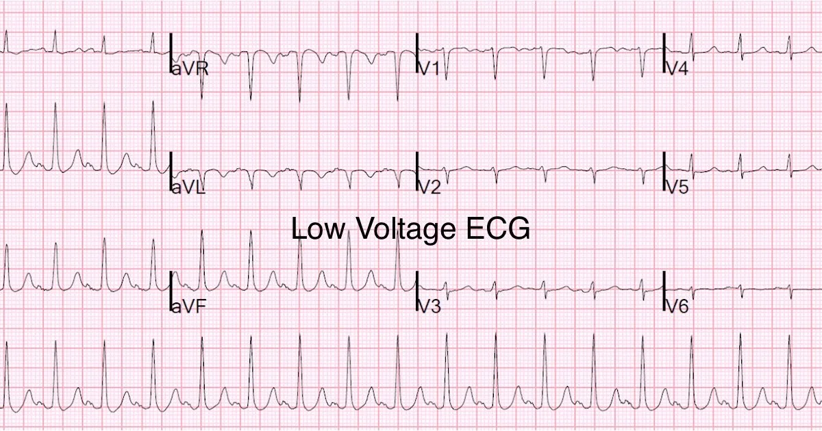
The Low Voltage ECG
A “low voltage,” ECG is a sign that something is getting in the way of the electrical conduction of the heart and the EKG leads on the patient’s chest.
Low voltage is concerning for a condition where fluid collects between the heart’s muscle and the thin protective sac around it (the pericardium), called a pericardial effusion.
Untreated severe pericardial effusion can eventually stop the heart from beating.
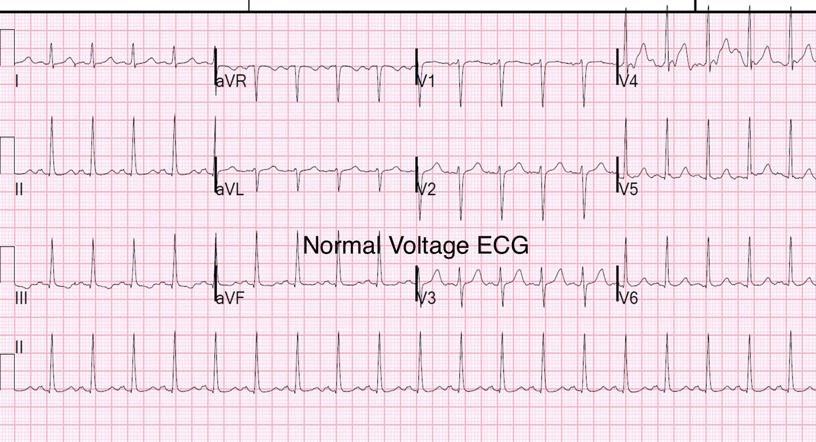
The QRS is said to be low voltage when:
The amplitudes of all the QRS complexes in the limb leads are < 5 mm; or
The amplitudes of all the QRS complexes in the precordial leads (V1-V6) are < 10 mm
Mechanisms
Low voltage is produced by:
The “damping” effect of increased layers of fluid, fat, or air between the heart and the recording electrode
Loss of viable myocardium
Diffuse infiltration or myxoedematous involvement of the heart
Causes
The most important cause is massive pericardial effusion, which produces a triad of:
Low voltage
Tachycardia
Electrical alternans
Patients with this triad need to be immediately assessed for clinical or echocardiographic evidence of tamponade.
Other causes of a low voltage ECG include:
Fluid:
Pericardial effusion; Pleural effusion
Fat:
Obesity
Air:
Emphysema; Pneumothorax
Infiltrative / Connective Tissue Disorders
Myxoedema
Infiltrative myocardial diseases —
i.e. restrictive cardiomyopathy due to amyloidosis, sarcoidosis, hemochromatosis
Constrictive pericarditis
Scleroderma
Loss of viable myocardium:
Previous massive MI
End-stage dilated cardiomyopathy